Posts

Showing posts with the label health

The eyes : ocular herpes simplex , ocular herpes zoster , ophthalmic examination , Physical Examination , Visual Acuity and Visual Field , Slit Lamp Examination , Tonometry , ophthalmoscopy , optical coherence tomography (OCT) , optic nerve , optic nerve atrophy and orbital cellulitis .

Image
ocular herpes simplex An INFECTION of the eyes with HERPES SIMPLEX VIRUS 1 (HSV-1), which causes cold sores, or herpes simplex virus 2 (HSV-2), which causes GENITAL HERPES. The virus spreads to the eye to cause the initial infection via contamination from contact with an existing herpes sore elsewhere on the body. Ocular herpes simplex features similar eruptions of sores on the surface of the EYE and inside the eyelids. The sores are very painful and can cause permanent scarring of the CORNEA. About half of people who have one outbreak of ocular herpes simplex will experience a second; about 20 percent have persistently recurring infections, ranking ocular herpes simplex as the leading infectious cause of corneal destruction. A serious complication of ocular herpes simplex is stromal KERATITIS, in which the IMMUNE SYSTEM begins to attack the stromal cells that make up the cornea. This leads to scarring deep within the cornea, resulting in distortions of vision and diminished VISUAL ...

The eyes : night blindness and nystagmus .

Image
nearsightedness See MYOPIA. night blindness Impaired dark adaptation resulting from slowed photochemical reactions in the rods, the specialized cells of the RETINA that perceive contrast and detect visual images in low light. Night blindness becomes increasingly common after middle age. The person with night blindness may be unable to see at all in dim light or may experience delayed adjustment when going from a lighted environment to a dim or dark  environment. A diminished VISUAL FIELD with restricted peripheral vision also contributes to night blindness, as the outer edge of the retina where peripheral vision takes place contains mostly rods. There are not many treatment options for night blindness. Nutritional supplementation of vitamin A and the antioxidants LUTEIN and ZEAXANTHIN, which some studies show help maintain the health of the eye and improve the functioning of the rods, seem to aid some people. Adequate lighting when reading and especially when watching televi...

The eyes : lens , mydriasis and myopia .

Image
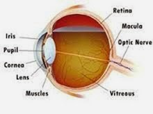
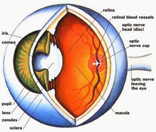
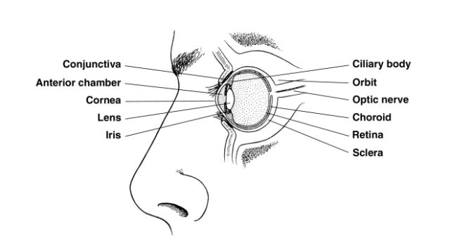
lens The primary focusing structure of the EYE, located in the center at the front of the eye. The lens is transparent, convex (rounded outward on each side), round, and flexible. A thin membrane encloses the lens. Tiny muscles at the front edges of its sides, the ciliary muscles, contract to flatten the lens and relax to thicken the lens. These adjustments alter light refraction (the angle at which the lens bends lightwaves entering the eye) to accommodate near and distant vision. The most common health conditions that affect the lens are PRESBYOPIA, in which the FLEXIBILITY of the lens diminishes with aging, and CATARACT, in which protein deposits cloud the lens and obscure vision. The lens is also vulnerable to accidental injury, particularly from blunt force (such as a baseball) or puncture. For further discussion of the lens within the context of ophthalmologic structure and function please see the overview section “The Eyes.” See also CATARACT EXTRACTION AND LENS REPLACE- ...

The eyes : keratitis and keratoconus

Image
keratitis INFLAMMATION of the CORNEA, usually the result of an INFECTION. The cause of the infection is more commonly viral, such as HERPES SIMPLEX or HERPES ZOSTER, than bacterial. Symptoms of infectious keratitis include • redness and irritation of the EYE and conjunctiva (inner eyelids) • discomfort or PAIN • excessive tearing • difficulty keeping the eye open • diminished VISUAL ACUITY (usually blurred vision) • eye discharge or crusting Viral keratitis usually runs its course without complication, though occasionally a secondary bacterial infection may develop. ANTIVIRAL MEDICA- TIONS sometimes shorten the course of chronic herpes infections. Bacterial keratitis typically follows a CORNEAL INJURY, such as an abrasion or laceration, and requires treatment with ophthalmic ANTIBIOTIC MEDICATIONS. Chronic or recurrent keratitis can cause permanent scarring of the cornea, resulting in diminished visual acuity such as ASTIG- MATISM. Extensive corneal damage may require C...

The eyes : intraocular pressure , iritis and ischemic optic neuropathy .

Image
intraocular pressure The pressure within the EYE that maintains the eye’s form and structure. Nor- mal intraocular pressure in an adult is 12 to 22 millimeters of mercury (mm Hg). A device called a tonometer measures intraocular pressure, either through light contact against the anesthetized eye or via the force of resistance to a puff of air blown against the eye’s surface (a noncontact method that does not require anesthetic drops). Elevated intraocular pressure is called ocular hypertension. Lower than normal intraocular pressure is called ocular hypotension. Ocular hypertension (greater than 21 mm Hg) is a hallmark sign of GLAUCOMA, an eye condition that, if untreated, results in complete loss of vision. Other health conditions that can increase intraocular pressure include tumors that press against the eye, ORBITAL CELLULITIS, and GRAVES’S OPHTHALMOPATHY. Increased intraocular pressure damages the cells on the front of the OPTIC NERVE (the retinal ganglia), leading to permane...

The eyes : hordeolum and hyperopia .

Image
hordeolum A bacterial INFECTION of a gland or an eyelash follicle along the edge of the eyelid, commonly known as a stye. A hordeolum causes swelling, redness, PAIN, and a discharge that leaves a crusty layer on the eyelids during sleep. The doctor can usually diagnose hordeolum by its presenta- tion. BLEPHARITIS (infection of the inside surface of the eyelid) and CONJUNCTIVITIS (infection of the conjunctiva, the membrane lining the eyelids) may instigate, accompany, or follow hordeolum. The doctor often chooses to anesthetize the area and lance (make tiny punctures or incisions under sterile conditions) the hordeolum to drain its contents and relieve the pressure. Further treatment is ophthalmic ANTIBIOTIC MEDICATIONS, typically in ointment form, applied to the area. Sometimes oral antibiotic medications are also necessary. Warm, moist compresses soothe the irritated tissues and help draw out any remaining pus. Most hordeola clear up in 7 to 10 days with treatment and heal withou...

The eyes : glaucoma , Symptoms and Diagnostic Path , Treatment Options and Outlook , Risk Factors and Preventive Measures , Graves’s ophthalmopathy , Symptoms and Diagnostic Path , Treatment Options and Outlook and Risk Factors and Preventive Measures

Image
glaucoma A serious and progressive EYE condition in which the cells at the front of the OPTIC NERVE where it intersects with the RETINA, the retinal ganglia, die, resulting in vision loss. Early diagnosis and treatment can minimize vision loss. Health experts estimate that 5 million Americans have glaucoma, though only about 2 million of them know it. Glaucoma is the third-leading cause of blindness in the United States, primarily because it remains undetected until damage to the optic NERVE becomes significant. Glaucoma becomes more common after age 65, though there is a congenital form that manifests in early child- hood (congenital glaucoma). Until the late 1990s ophthalmologists perceived glaucoma to be the exclusive consequence of increased pressure within the eye (INTRAOCULAR PRESSURE) that caused the death of retinal ganglia cells. Current understanding of the disease process of glaucoma affirms the death of retinal ganglia cells as the cause of damage to the optic nerve, tho...

The eyes : flashes , floaters and fluorescein staining

Image
farsightedness See HYPEROPIA. flashes Visual phantoms that appear as spots of light. An ophthalmologist should evaluate occurrences of flashes, as they can be symptoms of RETI- NAL DETACHMENT or other conditions affecting the RETINA. Flashes represent stimulation of the rods and cones, the cells of vision carpeting the retina, that occurs when the gelatinous fluid holding the retina in place (vitreous humor) moves across them. Because the NERVE signals these cells send to the BRAIN encode patterns of light, the brain interprets messages from them as light. A blow to the head that causes a person to “see stars” has similar effect when it is forceful enough to jostle the vitreous humor against the retina. Flashes may appear as multiple spots of light, “light showers,” or lightning-like streaks. The ophthalmologist typically performs a full OPHTHALMIC EXAMINATION to assess the integrity of the retina. Prompt treatment is necessary to intervene with a retinal tear or retinal detachm...

The eyes : ectropion , electroretinography , entropion , enucleation , episcleritis , exophthalmos , eye and eye strain

Image
ectropion Loss of elasticity or control of the eyelid, usually the lower eyelid, that causes it to sag away from the EYE. Ectropion allows tears to over- flow the lid rather than remaining in the eye. It also fails to protect the eye, and especially the CORNEA, permitting dryness and exposure to environmental particles that create irritation and possibly injury to the cornea and sclera (“white” of the eye). Common causes of ectropion include • aging • damage to the nerves that control the eyelids • CICATRICIAL PEMPHIGOID Ectropion is a common symptom of BELL’S PALSY, a temporary paralysis of one side of the face that results from INFLAMMATION of the seventh cranial NERVE (facial nerve), and also may accompany neurologic disorders such as PARKINSON’S DISEASE and MULTIPLE SCLEROSIS. With ectropion the eye feels irritated and scratchy. Tear production becomes excessive as the eye attempts to lubricate and protect itself, and tears typically run over the lip of the lid and ont...

The eyes : dacryocystitis , dacryostenosis , dark adaptation test , diplopia and dry eye syndrome .

Image
dac r y o c ystiti s INFLAMMATION of the lacrimal (tear) ducts, typically the nasolacrimal ducts in the corners of the EYE near the NOSE. Dacryocystitis develops when there is a blockage of the lacrimal duct, which may result from DACRYOSTENOSIS (narrowing of the lacrimal duct), INFECTION, or chronic irritation such as might occur with ALLERGIC RHINI- TIS or ALLERGIC CONJUNCTIVITIS. Dacryocystitis can be acute (of sudden onset) or chronic (recurrent or long-standing). It also can be congenital (the result of defects of the lacrimal gland and duct structures) or acquired. Most people who have acquired dacryocystitis are over age 65. Common symptoms include • redness and swelling between the eye and the bridge of the nose • rhinitis (runny nose) • PAIN • overflowing tears • FEVER when an infection is present The doctor can typically diagnose dacryocystitis based on its presentation. Dye tests, in which the doctor places a special dye in the eye and watches to see whether t...

The eyes : cataract , age-related cataracts , congenital cataracts , cataracts of diabetes , symptoms and diagnostic path , treatment options and outlook , risk factors and preventive measures , surgical procedure , phacoemulsification , extracapsular cataract extraction , lens replacement , risks and complications , outlook and lifestyle modifications , corneal transplantation , cornea donation , eyeglasses and contact lenses

Image
cataract Cloudiness and discoloration of the LENS. Cataracts become increasingly common with advancing age, affecting half of all people age 80 and older. Cataracts were once a leading cause of age-related blindness. Today ophthalmologists surgically remove cataracts and replace the lens with a prosthetic intraocular lens (IOL) that restores vision. Cataracts result from protein deposits that accumulate within the lens. These deposits disperse light in much the same way cracks in a window might splinter sunlight shining through. The fragmented light creates areas of accentuated bright- ness, causing the halos and sensitivity to lights at night. The opacity of the cataract interferes with the refractive function of the lens, causing blurry or hazy vision. The yellow or gray discoloration of the lens common with mature or “ripe” cataracts filters the lightwaves that enter the EYE, particularly affecting those in the spectrum of blue. The location of the cataract on the lens determin...